AsdocAI
Symptom Checker
Image Diagnosis
Posts
Symptom Checker
Image Diagnosis
Posts
К
Комп'ютерна томографія придаткових пазух носа
1 posts
56022-02 - Community Discussions
Posts
New Post
V
A rhinolith is not a polyp, but symptoms can be similar
Vasyl Meniuk
Contributor
Verified Doctor
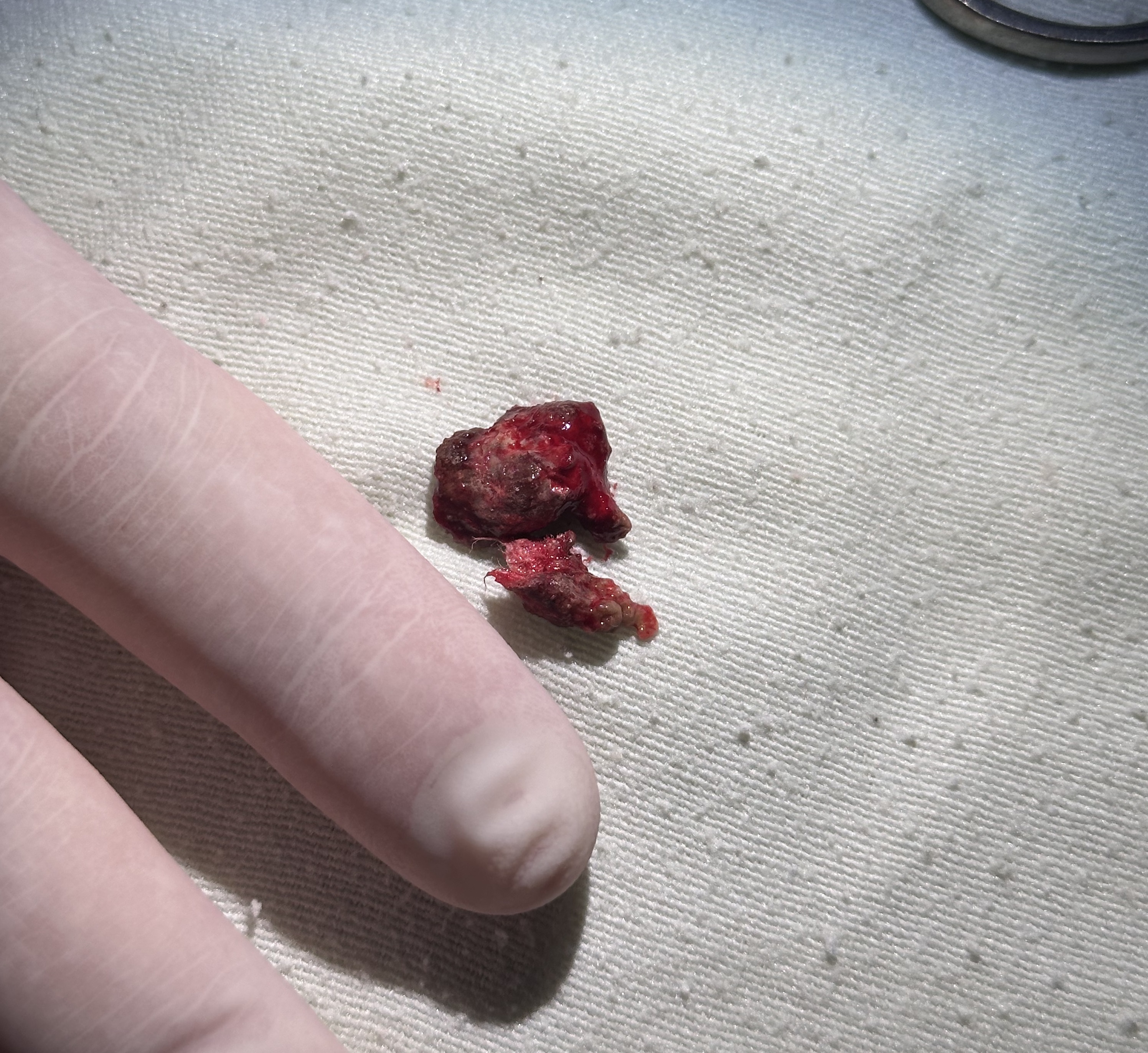
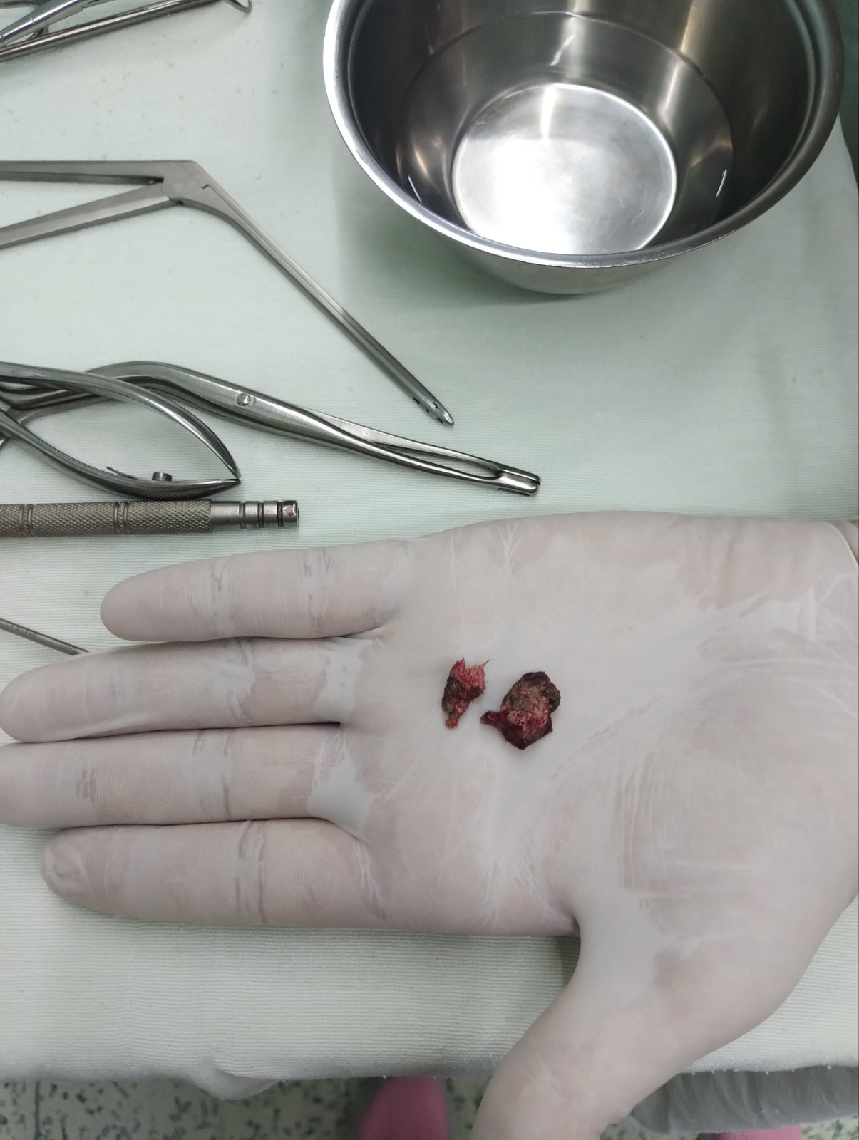
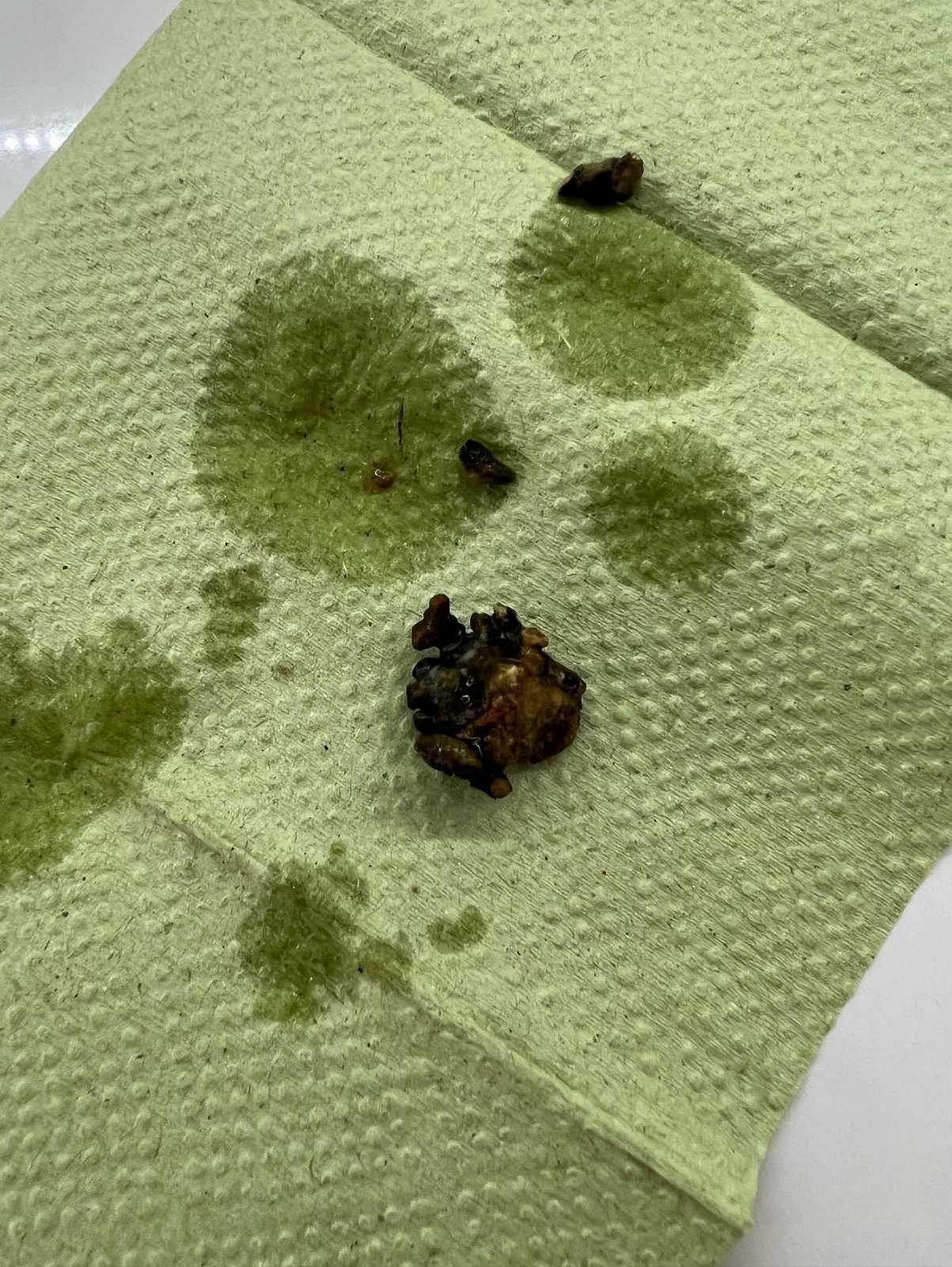
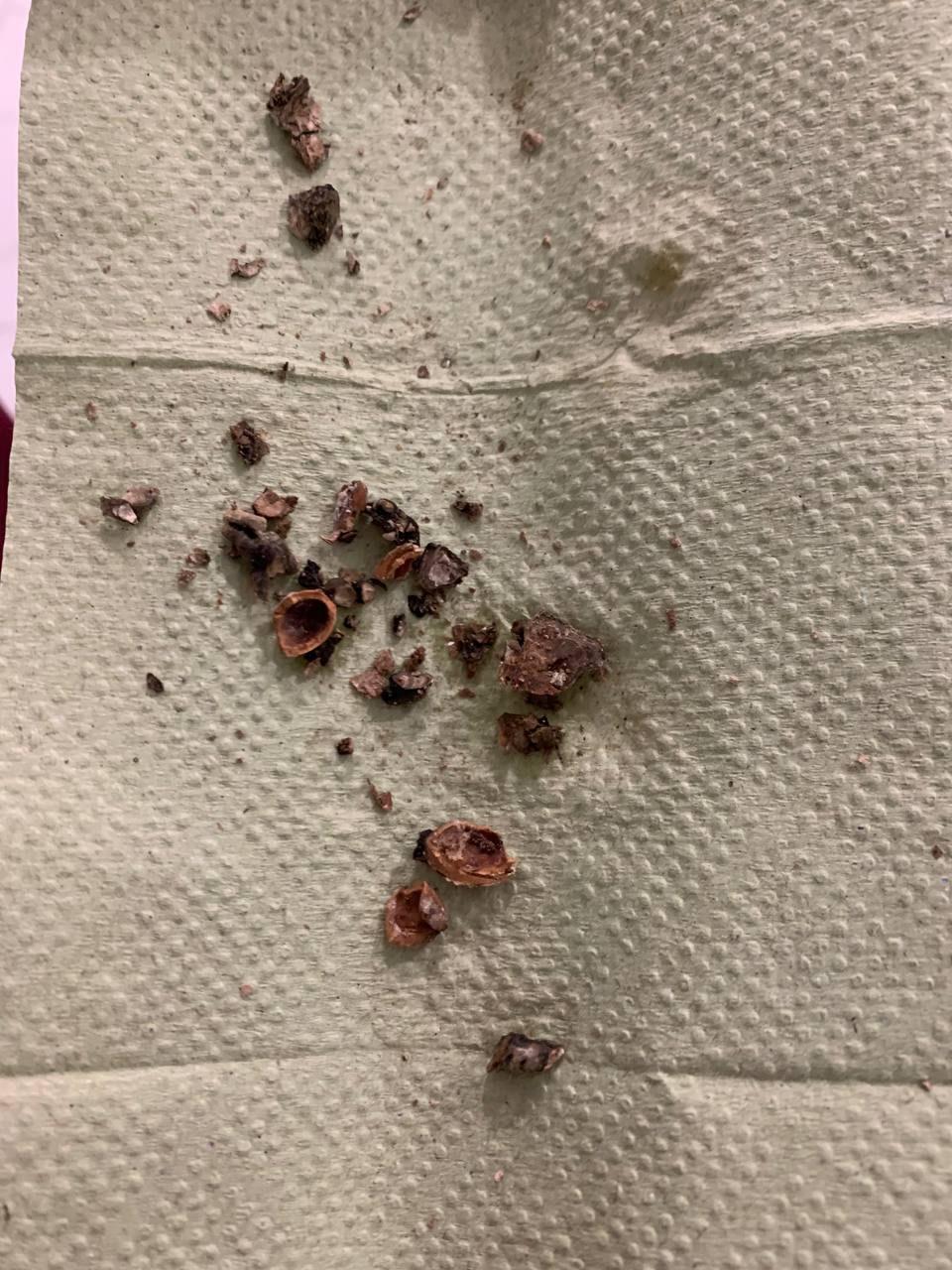
A rhinolith is a hard formation in the nasal cavity that forms over months or even years around a small foreign body, blood clot, or mucus due to the deposition of calcium salts. This is a rare condition that requires vigilance on the part of the otolaryngologist. It is often discovered incidentally during routine CT scans for dental or other reasons. Images clearly show a foreign object in the nasal passages or sinuses, or structural changes. Incidentally, regular use of head CT scans allows many ENT pathologies to be detected in their early stages, even before pronounced symptoms appear. Clinical case A 14-year-old female patient presented with complaints of nasal obstruction and persistent nasal discharge. She had undergone treatment multiple times, but the effect was transient. On examination following decongestion of the nasal passages, a mucus-covered foreign body was identified; it had a dense, chalky structure and was freely mobile, but could not be removed immediately. The patient was recommended to undergo a CT scan of the paranasal sinuses to clarify the structure and location of the rhinolith. It was subsequently successfully removed under general anesthesia. This is already the second case of a rhinolith in my practice—extremely interesting given the rarity of this condition. In the photos and video below, you can see the location of the rhinolith in the patient's nose, its CT appearance, and the removal process. Another case that may be of interest is also presented. If you have complaints regarding nasal breathing or other symptoms, do not delay—consult an otolaryngologist.
Риноліт
Виділення з носа
Стороннє тіло носа
Закладеність носа
Хронічний риніт (J31.0)
Стороннє тіло у носовому ході (T17.1)
Назальна ендоскопія (41764-00)
Комп'ютерна томографія придаткових пазух носа (56022-02)
1
0
4
6
Show Original